Electroencephalogram (EEG)
Introduction
Reference: Introduction to EEG
Standard Sensitivity: 7 µV/mm - 0.007 V/m
EEG - Origin and Measurement
![]() EEG-fMRI: Physiological Basis, Technique, and Applications (Mulert & Lemieux, 2023)
EEG-fMRI: Physiological Basis, Technique, and Applications (Mulert & Lemieux, 2023)
The EEG consists of the summed electrical activities of populations of neurons, with a modest contribution from glial cells. The neurons are excitable cells with characteristic intrinsic electrical properties, and their activity produces electrical and magnetic fields.
Two Types of Neuronal Activations
- Action Potential
- Synaptic Activation
- EPSP
- Positive ions inwards (e.g., \(Na^+\))
- IPSP
- Negative ions inwards (e.g., \(Cl^-\))
- Positive ions outwards (e.g., \(K^+\))
- EPSP
Sink-Source Configuration
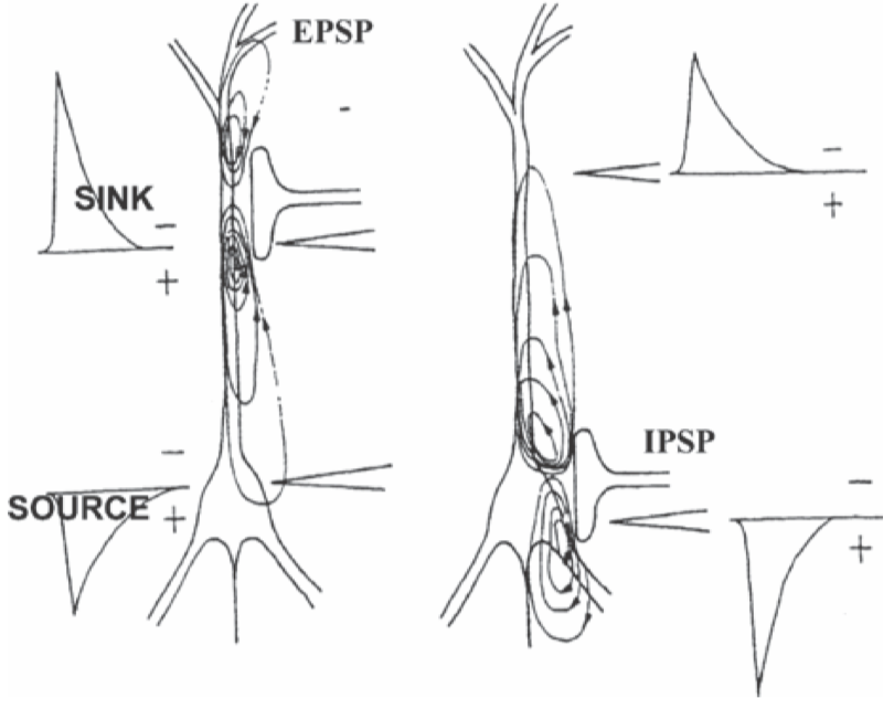
There is no accumulation of charge anywhere in the medium, the transmembrane currents that flow in or out of the neuron at the active synaptic sites are compensated by currents that flow in the opposite direction elsewhere along the neuronal membrane.
Active sink is generated in the extracellular medium at the level of an excitatory synapse. Distributed passive sources along the soma-dendritic membrane.
Active source is generated in the extracellular medium at the level of an inhibitory synapse. Distributed passive sinks along the soma-dendritic membrane.
The flows of these compensating extracellularly currents depend on the electrical properties of the local tissue. Glial cells occupy an important part of the space between neurons and are coupled to one another by gap junctions (the conductivity is sensitive to changes in \(pH\), extracellular \(K^+\) and \(Ca^{2+}\), and physiological/pathological conditions).
Reference
- Mulert, C., & Lemieux, L. (2023). EEG-fMRI: physiological basis, technique, and applications. Springer Nature.