Nerve Fiber
Types of Nerve Fiber
| Classification | Type | Subtype | Example | Relative Diameter | Myelination |
|---|---|---|---|---|---|
| Sensory & Motor |
A | \(\alpha\) | \(\alpha\) motor neurons | Large | Heavily myelinated |
| \(\beta\) | touch, pressure | Medium | Heavily myelinated | ||
| \(\gamma\) | intrafusal fibers | Medium | Heavily myelinated | ||
| \(\delta\) | touch, pressure, temperature, fast pain |
Small | Heavily myelinated | ||
| B | Small | Lightly myelinated | |||
| C | Small | Unmyelinated | |||
| Sensory | I | a | Muscle spindle afferents | Large | Myelinated |
| b | Golgi tendon organ afferents |
Large | Myelinated | ||
| II | secondary afferents of muscle spindles; touch, pressure |
Medium | Myelinated | ||
| III | touch, pressure, fast pain, temperature |
Small | Myelinated | ||
| IV | pain, temperature, olfactory |
Small | Unmyelinated |
Spinal Cord Nerve Fiber
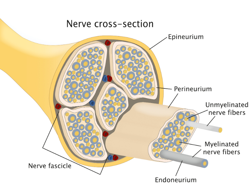 Schematic of the histologic structure of a peripheral nerve. Adapted from (Schmitd et al., 2021)
| Diameter(mm) | Number of nerve fibers | |||
|---|---|---|---|---|
| Segment | Ventral root (Motor) |
Dorsal root (Sensory) |
Ventral root (Motor) |
Dorsal root (Sensory) |
| C1 | 0.97±0.16 | 1.21±0.28 | 2751±639 | 6430±1606 |
| C2 | 1.34±0.30 | 2.61±0.51 | 3116±724 | 11947±2977 |
| C3 | 0.80±0.23 | 2.87±0.52 | 2460±471 | 21876±1916 |
| C4 | 1.39±0.24 | 2.49±0.34 | 3833±408 | 10647±887 |
| C5 | 2.50±0.55 | 3.43±0.77 | 7841±1020 | 23300±2856 |
| C6 | 2.23±0.73 | 3.99±0.75 | 7048±1157 | 36353±7451 |
| C7 | 2.22±0.50 | 4.61±0.87 | 8467±1019 | 39653±8458 |
| C8 | 1.71±0.60 | 3.92±0.62 | 5883±1000 | 31156±8273 |
| T1 | 1.03±0.23 | 2.18±0.31 | 5788±1186 | 26507±7617 |
| T2 | 0.75±0.11 | 1.30±0.14 | 3576±398 | 10234±1728 |
| T3 | 0.78±0.10 | 1.35±0.16 | 5499±1126 | 14888±2514 |
| T4 | 0.77±0.17 | 1.13±0.13 | 5485±973 | 10849±1832 |
| T5 | 0.64±0.08 | 1.21±0.30 | 5326±1314 | 8355±1390 |
| T6 | 0.71±0.18 | 1.07±0.16 | 3666±1407 | 10015±1666 |
| T7 | 0.78±0.15 | 1.25±0.27 | 4297±1130 | 9123±1178 |
| T8 | 0.83±0.25 | 1.29±0.18 | 3643±1340 | 7619±903 |
| T9 | 0.81±0.15 | 1.33±0.25 | 5209±704 | 8369±967 |
| T10 | 0.72±0.08 | 1.27±0.15 | 5269±963 | 11329±2724 |
| T11 | 0.69±0.08 | 1.26±0.16 | 4870±895 | 9713±1824 |
| T12 | 0.76±0.14 | 1.45±0.19 | 6538±892 | 10420±802 |
| L1 | 0.81±0.07 | 1.55±0.28 | 5384±833 | 16820±3456 |
| L2 | 0.96±0.16 | 1.93±0.27 | 7374±720 | 18615±±3284 |
| L3 | 1.19±0.07 | 2.24±0.30 | 9169±1160 | 26191±2772 |
| L4 | 1.04±0.07 | 2.48±0.38 | 7878±1386 | 31175±2686 |
| L5 | 1.37±0.16 | 2.66±0.40 | 8657±1396 | 34455±2740 |
| S1 | 1.43±0.16 | 2.95±0.57 | 8253±1419 | 41543±3036 |
| S2 | 0.93±0.11 | 2.02±0.53 | 4766±1035 | 18642±1716 |
| S3 | 0.55±0.07 | 1.32±0.60 | 2233±299 | 11971±964 |
| S4 | 0.34±0.03 | 0.52±0.17 | 1356±193 | 3402±304 |
| S5 | 0.14±0.02 | 0.27±0.13 | 906±111 | 2206±197 |
*Table adapted from (Liu et al., 2015).
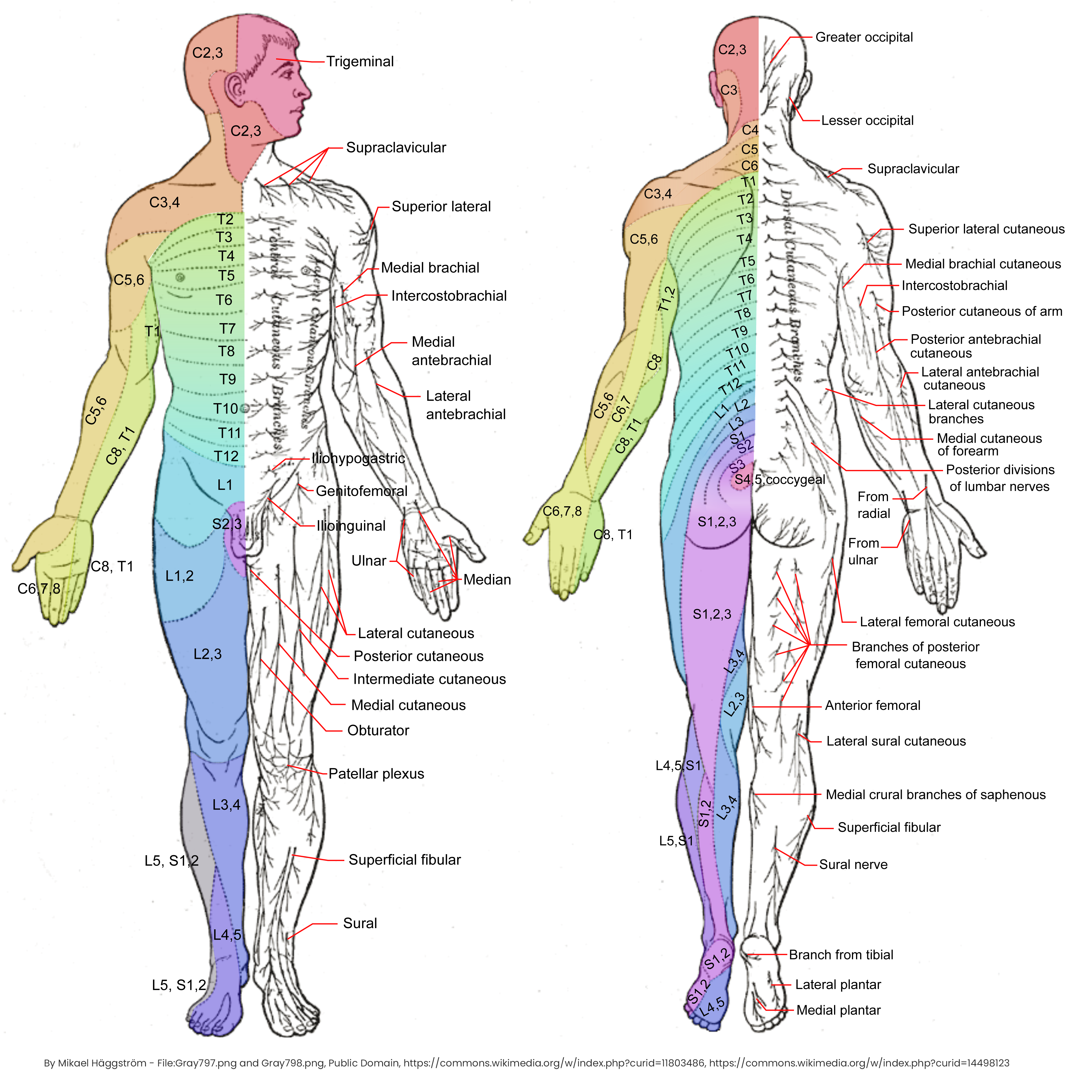 Dermatomes and Cutaneous Nerves (Wikipedia)
Optic Nerve Fibers
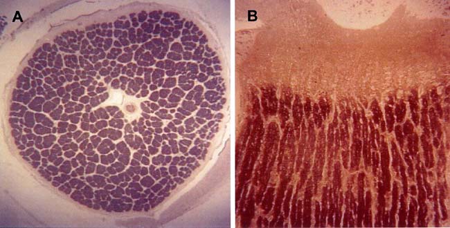 Image adapted from (Bose et al., 2005)
- Axon diameter: Mean=\(0.683 \mu m\), SD=\(0.369 \mu m\), SEM=\(0.026 \mu m\)
- Fig A: Normal human left optic never fiber cross-section (about 1million retinal ganglion cell axons).
- Fig B: Transverse section.
Miscellaneous
Lower Bound of Axon Diameter
Axon diameters are pushed toward the channel-noise limit of \(0.1 \mu m\) (mature neurons) due to Spontaneous Action Potentials (SAPs) (Faisal et al., 2005).
Thickness of the Cell Membrane
\(7.5 - 10 nm\) (Elert, 2023).
Depolarisation and Hyperpolarization Voltage
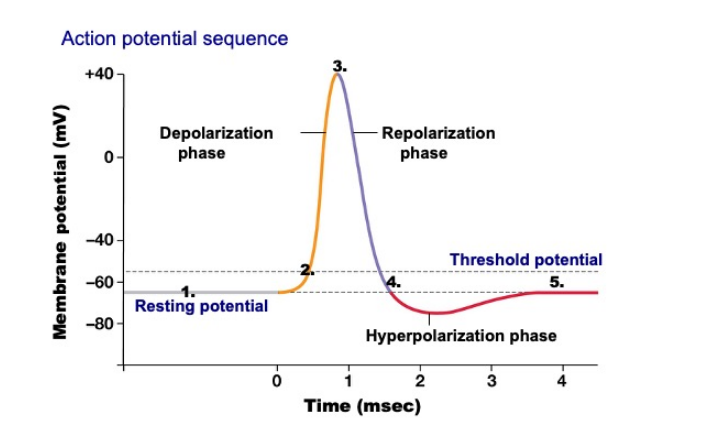
| Phase | Voltage | Duration |
|---|---|---|
| Depolarization | 110mv | 1ms |
| Repolarization | 110mv | 2~3ms |
| Hyperpolarization | 5mv | 1ms |
| Refractory Period | / | 2~3ms |
Firing Rate
From \(<1Hz\) to \(200Hz\) (minimum \(5ms/spike\)).
Axonal Conduction Velocity
From \(0.1m/s\) (unmyelinated) to \(200m/s\) (myelinated) (DeMaegd et al., 2017).
Endocytosis Size Limit
Max 500nm spheres in diameter - energy-dependent process (Rejman et al., 2004).
3nm Process
| Samsung | TSMC | |
|---|---|---|
| Transistor type | MBCFET | FinFET |
| Transistor density (MTr/mm2) | 150 | 197 (theoretical) 183 (A17 Pro) |
| SRAM bit-cell size (μm2) | Unknown | 0.0199 |
| Transistor gate pitch (nm) | 40 | 45 |
Transistor density: \(197\times 10^8 Tr/mm^2 = 197 \times 10^8 \div 1000^2 = 19700 Tr/{\mu m}^2\)
Cross-section area of a matured neuron (\(d=0.1 \mu m = 100 nm)\): \(3.14 \times 0.05^2 = 0.00785 {\mu m}^2\)
The number of transistors that can be fitted into the cross-section: 154 (upper bound)
Minimum transistor thickness: 0.34 nm (reference)
Size of multi-walled carbon nanotube: \(10-40nm, 5-20 \mu m\) (Abdallah et al., 2020).
Reference
- Abdallah, B., Elhissi, A. M. A., Ahmed, W., & Najlah, M. (2020). Carbon nanotubes drug delivery system for cancer treatment. In Advances in Medical and Surgical Engineering (pp. 313–332). Academic Press. https://doi.org/10.1016/B978-0-12-819712-7.00016-4
- Bose, S., Dhillon, N., Ross-Cisneros, F. N., & Carelli, V. (2005). Relative post-mortem sparing of afferent pupil fibers in a patient with 3460 Leber’s hereditary optic neuropathy. Graefe’s Archive for Clinical and Experimental Ophthalmology, 243, 1175–1179.
- DeMaegd, M. L., Städele, C., & Stein, W. (2017). Axonal conduction velocity measurement. Bio-Protocol, 7(5), e2152–e2152.
- Elert, G. (2023). The Physics Factbook. https://hypertextbook.com/facts/2001/JenniferShloming.shtml
- Faisal, A. A., White, J. A., & Laughlin, S. B. (2005). Ion-channel noise places limits on the miniaturization of the brain’s wiring. Curr. Biol., 15(12), 1143–1149. https://doi.org/10.1016/j.cub.2005.05.056
- Liu, Y. T., Zhou, X. J., Ma, J., Ge, Y. B., & Cao, X. (2015). The diameters and number of nerve fibers in spinal nerve roots. J. Spinal Cord Med., 38(4), 532. https://doi.org/10.1179/1079026814Z.000000000273
- Rejman, J., Oberle, V., Zuhorn, I. S., & Hoekstra, D. (2004). Size-dependent internalization of particles via the pathways of clathrin- and caveolae-mediated endocytosis. Biochem. J., 377(Pt 1), 159. https://doi.org/10.1042/BJ20031253
- Schmitd, L. B., Perez-Pacheco, C., & D’Silva, N. J. (2021). Nerve density in cancer: Less is better. FASEB BioAdvances, 3(10), 773.